New Study Challenges Assumptions about How Memories Are Made
TEHRAN (Tasnim) - New research reveals that short-term memories and long-term memories are formed simultaneously in different parts of the brain, a discovery that challenges our current models of memory formation.
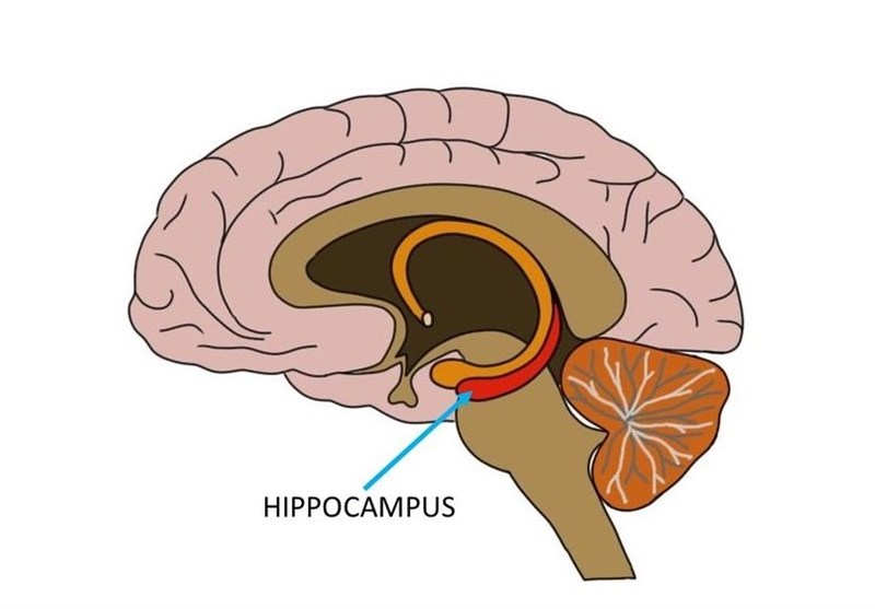
Until recently, scientists believed that memories were first stored in the hippocampus then gradually “transferred” to the cortex for long-term storage.
However, a new study conducted by the Riken-MIT Center for Neural Circuit Genetics challenges this theory, suggesting that memories are simultaneously stored in the hippocampus and cortex, but remain “silent” in the cortex for about two weeks before reaching a mature state.
Scientists have assumed that the engram cells in the cortex that store memory were generated slowly. The cells actually develop rapidly, said Takashi Kitamura, a lead author of the study, which is published in the journal Science, the Seker news website reported on Monday.
“Our study will partially revise current theory for memory consolidation,” Kitamura said. “Our theory for memory consolidation is that rapid generation and slow maturation of cortical engram cells are crucial for permanent memory storage in the cortex.”
The past theory that short-term memories in the hippocampus transfer to long-term memories in the cortex became was developed in the 1950s, when a famous amnesiac patient named Henry Molaison experienced a damaged hippocampus following surgery for epilepsy. He lost his ability to make new memories, but his long-term memories remained.
Until recently, scientists lacked a reliable way to test memory consolidation theory, Kitamura explained. “Most previous studies of memory were based on analyzing how damage to certain brain areas affects memory,” he said.
In 2012, when Tonegawa Laboratory, a neuroscience research lab at MIT, developed a way to label engram cells, this opened the door to new ways to test memory consolidation theories. “This method allows us to trace the circuits involved in memory storage and retrieval,” Kitamura said.
Kitamura and his team artificially reactivated memories in mice by using optogenetics, a technique that involves beaming light into their brains to control the activity of individual neurons by switching memories on or off. Researchers used this approach to label memory cells during a fear-conditioning event that involved a mild electric shock to the mouse and then used light to artificially reactive memories at different times.
Memory cells were labeled in three parts of the brain: the hippocampus, the prefrontal cortex and the basolateral amygdala, which stores memories’ emotional associations.
A day after the mice received an electric shock, there was evidence that memories of the fear-inducing event were stored in engram cells in both the hippocampus and the prefrontal cortex. The engram cells in the prefrontal cortex, however, remained silent. These cells stimulated freezing behavior when artificially activated by light, but did not fire during natural memory recall. Furthermore, when the connection between the hippocampus and cortex was blocked, long-term memory was unable to mature.
As a result of the study, researchers discovered that memory engram cells naturally change their status from active to silent and from silent to active. “If we identify the mechanisms behind how silent memory can be active, we may find a treatment for amnesia,” Kitamura said.
On the other hand, if researchers can identify the mechanisms behind how overactive memory can become silent, they may find a way to inhibit overly strong, negative memories. A better understanding of this process could potentially lead to new ways to treat post-traumatic stress disorder.